
The electrical activity of the heart is captured by an electrocardiogram. It is a routine test that is painless and used to swiftly identify heart issues and keep track of the heart’s health. It is a heart electrogram, which uses electrodes applied to the skin to create a graph of voltage vs time for the electrical activity of the heart. Each cardiac cycle results in the depolarization and repolarization of the cardiac muscle, which is detected by these electrodes (heartbeat). Numerous cardiac disorders, including irregular heartbeat (such as atrial fibrillation and ventricular tachycardia), insufficient coronary artery blood flow (such as myocardial ischemia and myocardial infarction), and electrolyte problems, result in changes in the normal ECG pattern (such as hypokalemia and hyperkalemia).
The P wave, which indicates depolarization of the atria, the QRS complex, which shows depolarization of the ventricles, and the T wave, which represents repolarization of the ventricles, are the three primary elements of an ECG.
Arrhythmia
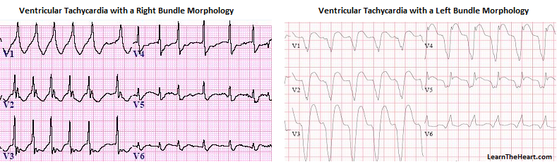
An arrhythmia is an abnormal heart rhythm. Some arrhythmias can cause problems with contractions of your heart chambers by: Not allowing the lower chambers (ventricles) to fill with enough blood, because an abnormal electrical signal is causing your heart to pump too fast or too slow. Not allowing enough blood to be pumped out to your body, because an abnormal electrical signal is causing your heart to pump too slowly or irregularly. Not allowing the top chambers (atria) to squeeze correctly. An arrhythmia can occur in the sinus node, the atria, or the atrioventricular (AV) node. These are called supraventricular arrhythmias. Arrhythmias can also occur in your ventricles and are caused by an abnormal electrical focus within your ventricles. This results in abnormal conduction of electrical signals within your ventricles. Arrhythmias can also be listed as slow (bradyarrhythmia) or fast (tachyarrhythmia). “Brady-” means slow, and “tachy-” means fast.

Heart Attack
Anterior ST Segment Elevation MI This is the big one that carries a high mortality if not treated rapidly. An anterior STEMI is usually from acute thrombotic occlusion of the left anterior descending coronary artery — also known as the “widow maker.”
QRS complex: This is the large peak that appears on a heart wave. The ventricles cause this wave when they pump blood out of your heart. ST-segment: This is a short section immediately after the QRS complex. Normally, there shouldn’t be any electrical activity in that segment, causing it to be flat and back to baseline. When there’s an elevation in the ST segment, that often means there’s a total blockage of one of the heart’s main supply arteries. When that is happening during a heart attack, it can be a sign that the muscle of the ventricles is dying. That’s critical information for healthcare providers to know during a STEMI because it means the heart muscle is in the process of dying. That also means reopening that artery and restoring blood flow as soon as possible may prevent permanent damage, or at least limit the severity of the damage.

Cardiomyopathy
Hypertrophic obstructive cardiomyopathy is a pathologic cardiac condition in which the interventricular septum is abnormally thickened. The classic ECG finding in hypertrophic obstructive cardiomyopathy is large dagger-like “septal Q waves” in the lateral — and sometimes inferior — leads due to the abnormally hypertrophied interventricular septum. Criteria for left ventricular hypertrophy is usually present. Wolff-Parkinson-White, or WPW, syndrome can be associated with HOCM as well.

Coronary Heart Disease
CAD is when a buildup of fatty substances blocks or interrupts the heart’s blood supply.
A normal ECG reading shows a consistent pattern across the P, QRS, and T waves. In a normal reading, both the ST segment and the T wave shows no signs of flattening, sharp spikes, or depressions. By contrast, an ECG reading of a severely diseased heart is noticeably different. The T-waves may flatten or have more of a downward slope, while the ST segments may have abnormal elevations or depressions, for example.

References.
1.https://ars.els-cdn.com/content/image/1-s2.0-S0950705117302769-gr2.jpg
5.https://my.clevelandclinic.org/health/diseases/23349-bradyarrhythmia